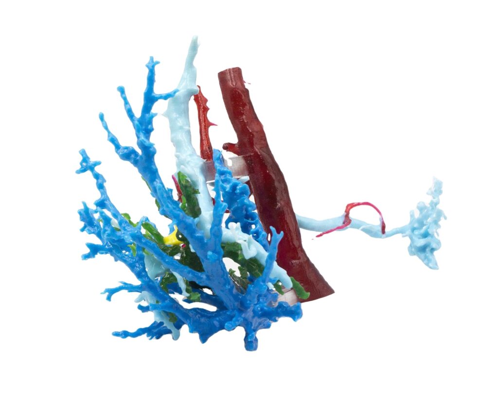
3D printing technology is a new type of rapid prototyping technology that builds objects layer by layer using biological materials extracted from metals, ceramics, polymers and composite materials that can be three-dimensionally printed, based on digital models. In neurosurgery, after obtaining CT, MRI, CTA, DSA and other imaging data, through software processing, 3D segmentation of target structures (such as skull, cerebrovascular, tumor, etc.) is realized, and the model is finally printed out with a 3D printer. Show the details of the patient’s skull.
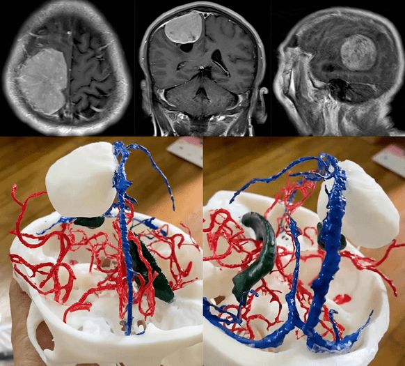
Case 1: was a giant meningioma at the top of the forehead. Due to the huge size of the tumor, and the anatomical structures adjacent to the tumor were displaced and deformed to varying degrees, the trainees used traditional normal anatomical teaching aids and planar imaging. The data is difficult to form a complete 3D imaginary space in a pathological state, and it is impossible to accurately identify the adjacent anatomical structures involved.
The model constructed by 3D printing technology intuitively and accurately restores the local anatomical structure of the tumor, allowing students to better understand the relationship between the tumor and the blood supply arteries, sagittal sinus, and the ventricular system affected by the tumor’s anatomical position changes. Information such as location and shape.
Case 2: was an unruptured posterior communicating aneurysm. Because the surgical planning of intracranial aneurysm involves the shape and size of the aneurysm, the width of the aneurysm neck, and the orientation of the aneurysm, many factors such as the shape and size of the aneurysm and the orientation of the aneurysm are affected. principle. Although 3D reconstruction can be achieved through the imaging data of the DSA examination in clinical practice, the students said that due to the fixed viewing angle of the 3D image of DSA and the inability to display the bone structure, there are still certain difficulties in the process of understanding.
Through the 3D printing model, students can observe the shape and size of the aneurysm and the relationship with the artery bearing the aneurysm from various angles. At the same time, they can also understand the anatomical position relationship between the aneurysm and the skeletal structure of the skull base, so as to better understand the artery. Surgical plan for tumor clipping.
